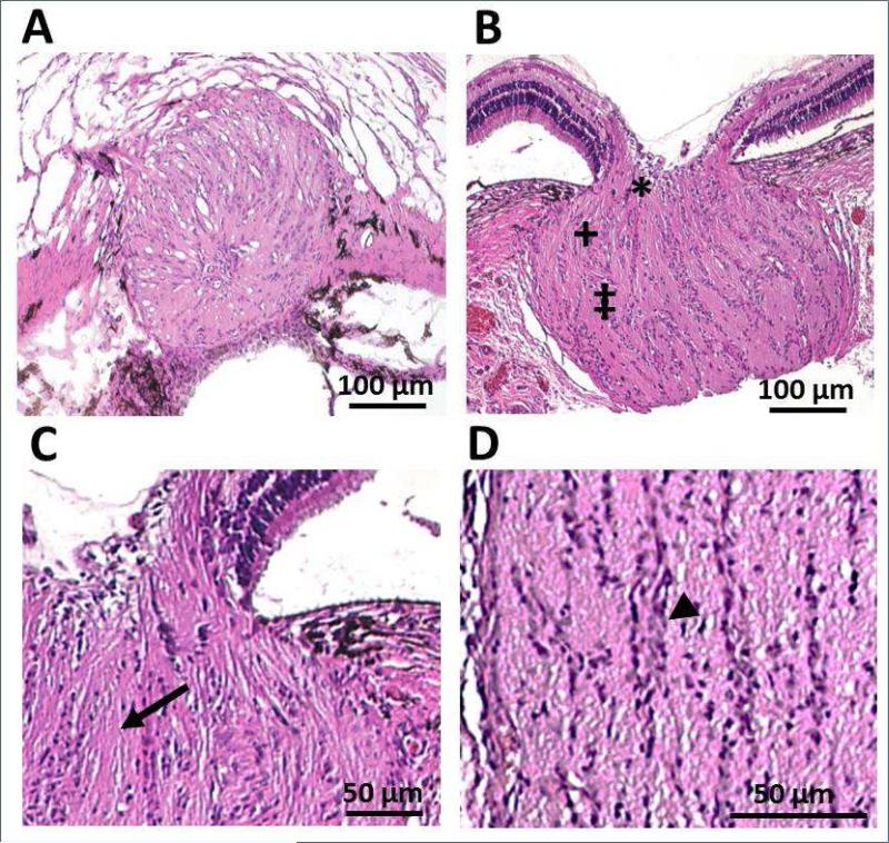
Figure 4.
H & E stained sections of the optic nerve head of a 9 weeks-old pigmented guinea pig; low magnification A) en face view at the level of the lamina cribrosa, cut at a slight angle with visible sclera superiorly and retina inferiorly, and B) cross-sectional view, including pre-laminar (*), laminar (+) and post-laminar regions (‡). Laminar region represents the section shown in (A); high magnification cross sectional views of C) pre-laminar and laminar regions (arrow: nerve fiber bundles) and D) the post-laminar region (arrowhead: presumed glial cells). Scale bars represent 100 μm (A & B) and 50 μm (C & D).

