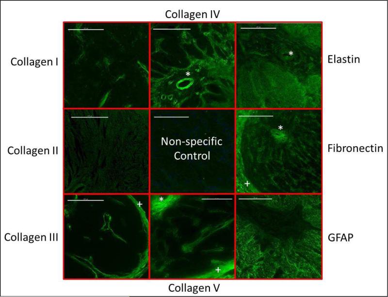
Figure 5.
Immunostained sections of the optic nerve head of a pigmented guinea pig, taken at the level of the lamina, showing the presence of collagen types I, III IV and V, as well as elastin, fibronectin and GFAP. Collagen II was not found in this region. Scale bar represent 100 μm. * vascular sheath; + surrounding meninges

