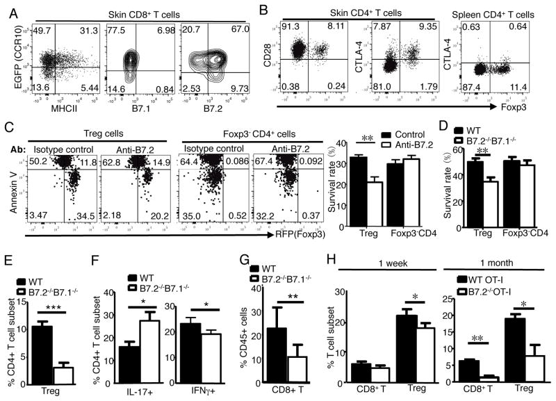
Figure 4.
Resident CD8+ T cells support maintenance of Treg cells in the skin through B7.2/receptor axis. A) Expression of MHCII, B7.1 and B7.2 on skin CD8+ T cells. One representative of 4 analyses of 2 experiments. B) Expression of CD28 and CTLA-4 on skin and splenic Treg and CD4+ Teff cells. Representative of 4 analyses. C) FC analysis of survival of WT Treg and Foxp3− CD4+ Teff cells 1 day after co-culture with WT CD8+ T cells in presence of anti-B7.2 or isotype control antibodies. Bar graphs show percentages of live Annexin V− RFP+ Treg and Annexin V− Teff cells. N=6 pooled of 2 experiments. D) Survival of WT Treg and Foxp3− CD4+ Teff cells 1 day after co-culture with WT or B7.1−/−B7.2−/− CD8+ T cells, performed same as in (C). N=4 pooled of 2 experiments. E–G) Percentages of Treg cells (E), IL-17+ and IFNγ+ CD4+ cells (F) and CD8+ cells (G) in the skin of Rag1−/− mice 1 month after they are transferred with WT or B7.2−/−B7.1−/− CD8+ and WT CD4+ T cells. N=5 pooled of 2 experiments. H) Analysis of WT and B7.2−/− donor OT-I cells and host Treg cells in the torso skin of WT recipients 1 week and 1 month after Ova immunization on ears. N=3 each.

