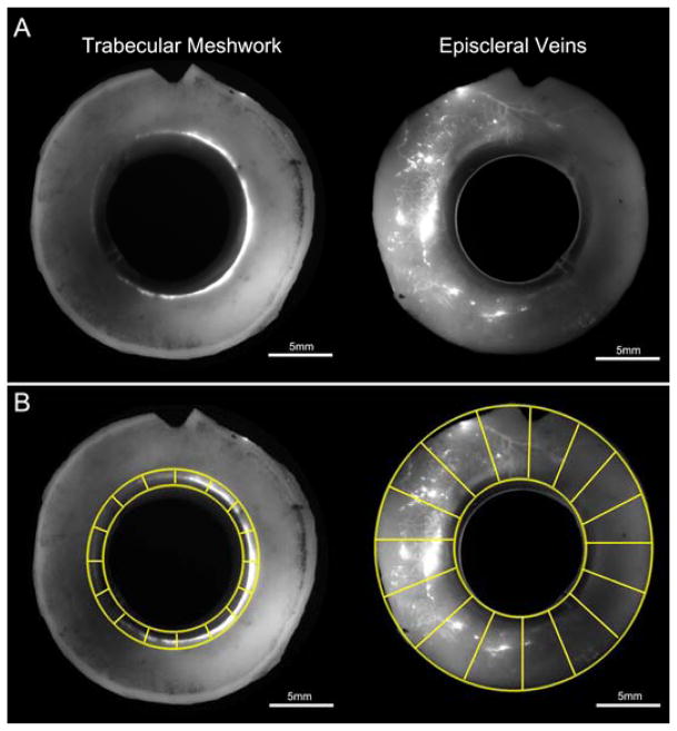
Figure 1. Global Imaging of Normal Human Eyes.
A) Global image of anterior segments from a pair of normal human eyes that reveals fluorescent tracer patterns in the trabecular meshwork and episcleral veins. B) Digital separation of global images of the trabecular meshwork and episcleral veins into a minimum of 16 “wedges”.

