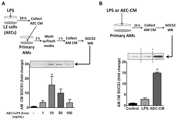
Figure 3. LPS induces elaboration of AEC-derived factors that can enhance SOCS3 secretion by AMs in vitro.
(A) AECs were plated at a concentration of 5 × 10⁵ cells/well of a 6-well plate and equilibrated for 16 h. The cells were washed and cultured with the indicated doses of LPS in serum-free RPMI for 24 h and the resulting AEC-CM was collected and cleared of cellular debris and apoptotic bodies. Primary AMs were collected and adhered for 1 h. The AMs were washed and incubated with AEC-CM for 2 h. After a second wash the AMs were cultured in serum-free RPMI for an additional 1-h and the resulting AM-CM was collected and analyzed for SOCS3 protein by WB. (Top) Schematic of experiments in which CM from AECs treated with LPS is incubated with AMs for subsequent evaluation of SOCS3 secretion. (Bottom) AM SOCS3 secretion by western blot analysis; above, representative blot; below, fold increase in SOCS3 compared to control based on densitometric analysis. (B) Direct versus indirect effect of LPS on AM SOCS3 secretion. 1×10⁶ AMs were adhered and incubated for 24 h in serum-free RPMI with either 10 ng/mL LPS or with CM from AECs that had been incubated for 24 h with 10 ng/mL LPS. The CM was collected as previously described and analyzed for SOCS3 by WB; above, representative blot; vertical dashed line indicates that the two lanes depicted were from the same blot but were not contiguous; below, fold increase in SOCS3 compared to control based on densitometric analysis. Data are expressed as mean ± SEM from three separate experiments, *= p<0.05.

