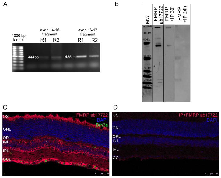
Figure 1. FMRP is expressed in the mouse retina under a 12h light/12h dark cycle.
A) Ethidium bromide-stained agarose gel with PCR reactions from two different mouse retinas (R1 and R2). Bands represent FMRP mRNA fragments from exon 14 to exon 16 (444 bp) and from exon 16 to exon 17 (435 bp). Bolder band in the ladder is 500 bp. B) Western blots of mouse retina protein samples with the ab17722 FMRP antibody. It labels bands at the expected 75 kDa size, but also one band at approximately 210 kDa (FMRP ab17722). Pre-adsorbing the antibody with an inhibitory peptide for 30 min (FMRP +IP 30′) or 24h (FMRP +IP 24h) blocks the labeling. C) Immunostaining of the mouse retina with the ab17722 FMRP antibody (red), cell nuclei are labeled with DAPI (blue). Labeling appears mostly in the OS, OPL, INL, IPL and GCL (two independent experiments from three animals). D) Pre-adsorbing the antibody with an inhibitory peptide makes the staining almost disappear (two independent experiments from three animals). OS: photoreceptor outer segments, OPL: outer plexiform layer, INL: inner nuclear layer, IPL, inner plexiform layer, GCL: ganglion cell layer.

