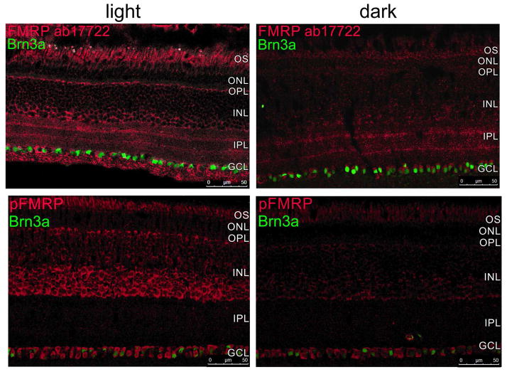
Figure 7. FMRP expression is modulated mostly in the OPL, INL and IPL in the chick retina.
Double-labeling immunohistochemistry of the ganglion cell marker, Brn3a (green) and FMRP (ab17722, red, top) and S499 phosphorylated FMRP (pFMRP, red, bottom panels) antibodies show that FMRP is located in the OS, OPL, INL, IPL and GCL (both in ganglion cells labelled with Brn3a and displaced amacrine cells negative for Brn3a ganglion cell marker). OS staining was considered unspecific because Fmr1 knockout retinas presented OS staining as well. It is noticeable that FMRP and pFMRP staining is stronger in light adapted compared to dark adapted retinas, except for the cells in the GCL. Three independent experiments were performed from three animals in each group.

