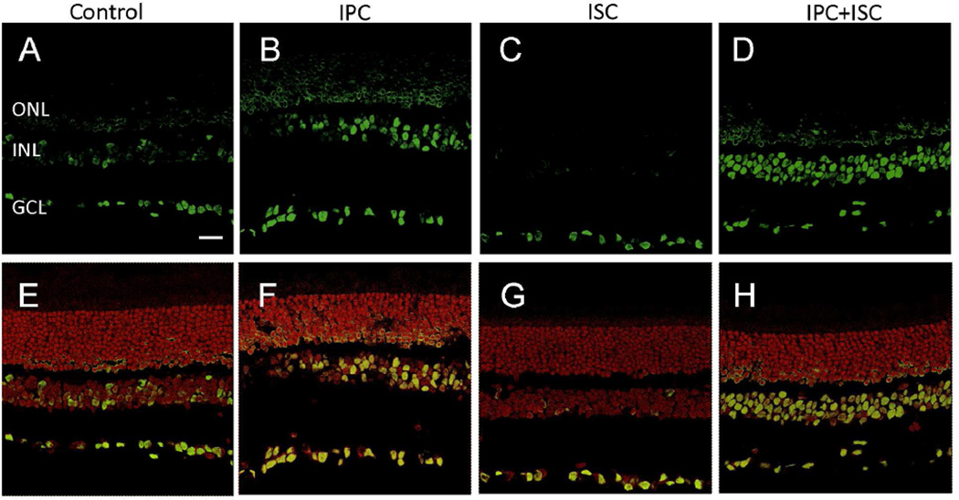
Figure 3.
Effect of IPC on acetylated histone-H3 immunostaining in rat retina cross sections. Immunohistochemical staining of acetylated histone-H3 (green) in (A) control, (B) IPC, (C) ischemia, and (D) IPC plus ischemia retinas; and sections overlaid with propidium iodide, nuclei staining (red) in (E) control, (F) IPC, (G) ischemia, and (H) IPC plus ischemia retinas. ONL, outer nuclear layer; INL, inner nuclear layer; GCL, ganglion cell layer. Scale bar: 20 µm.

