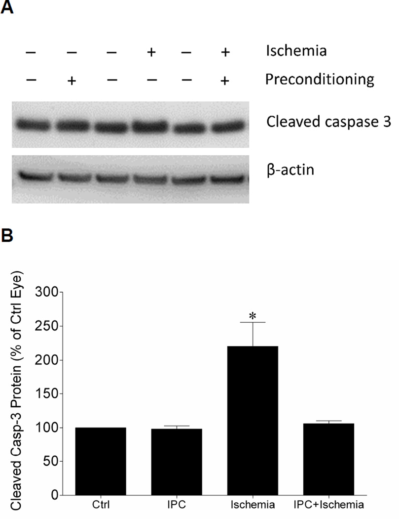
Figure 4.
Effect of IPC on retinal cleaved caspase-3 levels (A) Representative Western blot of retinal lysates for cleaved caspase-3 and β-actin at 24 hours after initiation of ischemic injury. (B) Levels are expressed as a mean percentage of the IPC or ischemic eyes relative to the control eyes. Data are expressed as mean ± SE, n=4. *Indicates significant difference (P <0.05), n=4.

