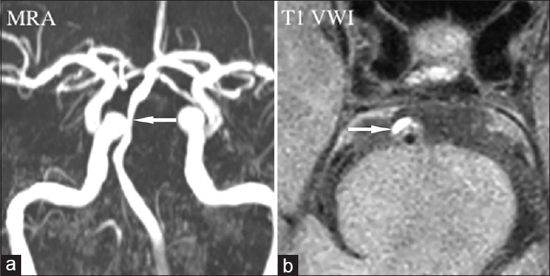
Figure 1.

Atherosclerotic stenosis. Magnetic resonance angiography (a) showed luminal stenosis in basilar artery (arrow). T1-weighted vessel wall image displayed the eccentric plaque locating in the ventral wall with high signal indicating intraplaque hemorrhage (b, arrow). MRA: Magnetic resonance angiography; VWI: Vessel wall imaging.
