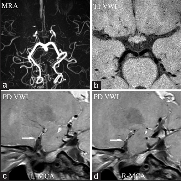
Figure 4.

Moyamoya disease. Magnetic resonance angiography (a) showed luminal occlusion in bilateral anterior cerebral artery and middle cerebral artery, suggesting possible Moyamoya disease. Normal vessel structure of bilateral anterior cerebral artery and middle cerebral artery disappeared in T1-weighted vessel wall image (b). Short-axis view of proton density-weighted vessel wall image displayed shrinking middle cerebral artery (c, arrow), especially in the right side (d, arrow). MRA: Magnetic resonance angiography; VWI: Vessel wall imagin; PD VWI: Proton density VWI; L: Left; R: Right; MCA: Middle cerebral arteries.
