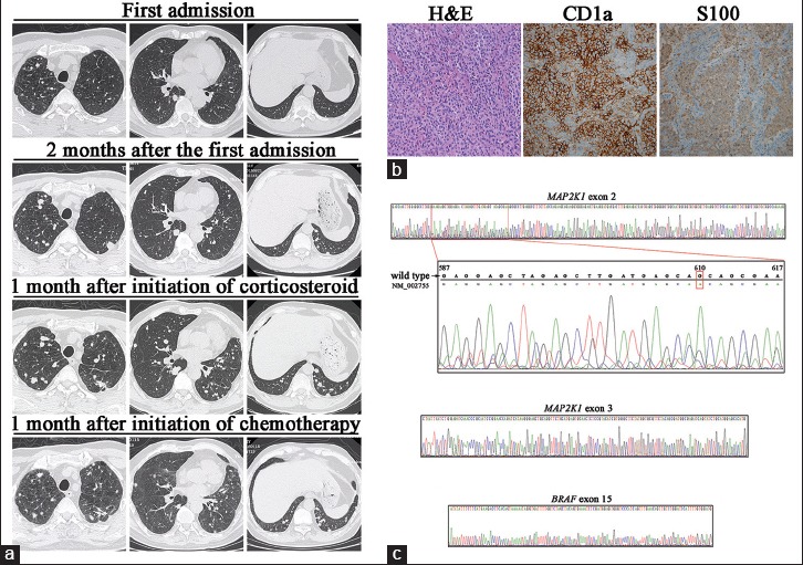
Figure 1.

(a) Chest high-resolution computed tomography findings of the patient. (b) Histopathological findings of the lung biopsy specimen. H and E stain showed cellular nodule with aggregates of Langerhans cells (original magnification ×200); immunohistochemistry for CD1a and S100 demonstrated positive staining in Langerhans cells (original magnification ×200). (c) Sequencing of MAP2K1 and BRAF. One silent mutation (code “CAG” to “CAA” at position 610) in exon 2 of MAP2K1 was detected while no mutations in exon 3 of MAP2K1 or in exon 15 of BRAF were detected.
