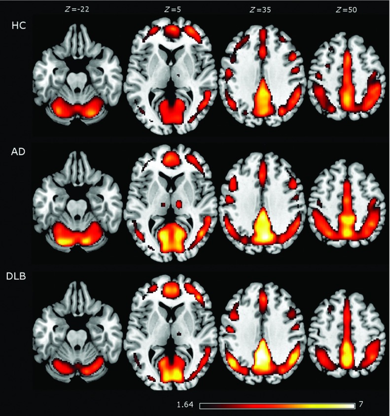
Figure 1.
The average regional homogeneity (ReHo) from healthy controls (HCs), Alzheimer's disease (AD), and dementia with Lewy body (DLB) patients. The highest ReHo values were found in the precuneus and frontal cortices in the three groups. Average images are shown with a t-score of >1.64, which is the threshold for the HC group when corrected for multiple comparisons (p-value of <0.05 corrected). Brain slices are shown in MNI standard space and in neurological convention (the left hemisphere is shown at the left).

