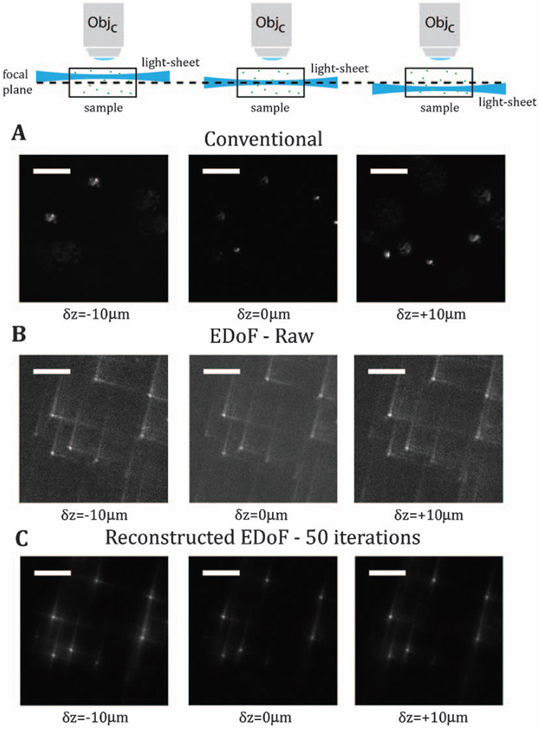
Fig. 2.
With conventional light-sheet microscopy, the tilt of the scanning beam results in out-of-focus imaging and loss of contrast of the scene in the focal plane. Panel A experimentally captures this loss of information through deterministic axial shifts of the scan beam as a controlled proxy for beam tilt. Panel B gives examples of the native images in the EDoF light-sheet system. Panel C demonstrates the results from deconvolving these images. The scale bar is 20 µm.

