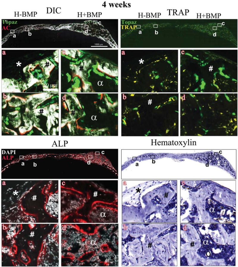
FIGURE 3.

Histology showing DIC-, TRAP-, ALP-, and hematoxylin-stained sections of the critical sized defects after 4 weeks of implantation with Healos with or without rhBMP-2 Full calvarial sections (scale bars: 1000 μm), magnified images a–d (scale bar: 50 μm). DIC sections show activity of GFP reporters in the regenerated tissues colocalized with alizarin complexone (AC). Bright green cells lined with a red mineralizing AC label are mature osteoblasts. Light green elongated cells form a periosteum-like layer at the periphery of the defects. TRAP: Bright yellow stain colocalized with light green cells represents TRAP positive host-derived osteoclasts. ALP: Red color indicates ALP positive cells. Cell nuclei are stained white with DAPI. (*) Host-derived fibroblastic periosteum-like layer; (#) trabecular-like bone with embedded osteocytes; (α) marrow-like matrix; and (circle) adipocytes. [Color figure can be viewed in the online issue, which is available at wileyonlinelibrary.com.]
