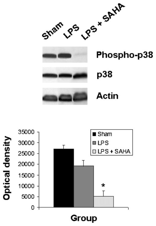
FIG. 1.
SAHA reduces the expression of phospho-p38. The cytosol fraction from the livers of mice treated with or without SAHA at 3 h after LPS insult was subjected to western blot with anti-phospho-p38, p38, and actin antibodies. Upper panel shows a representative western blot. Protein bands of phospho-p38 were quantified by densitometry and expressed as mean ± SD, n = 3. The asterisk indicates that a value significantly differs from the LPS group (P < 0.004). There was no difference between groups in the expression of non-phosphorylated p38.

