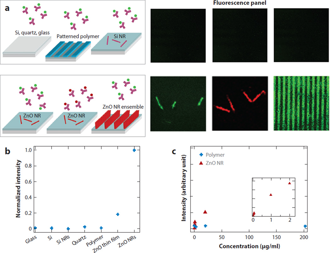
Figure 1.
Comparison of fluorescence emission monitored from 200-µg/ml fluorescein isothiocyanate (FITC)-conjugated or tetramethyl rhodamine isothiocyanate (TRITC)-conjugated antibovine immunoglobulin G on ZnO nanorods (NRs) versus control substrates after identical biodeposition. (a) No discernable fluorescence signal is detected from the biomolecules on the control substrates, including glass, quartz, native SiO2/Si, Si NRs, and polymeric surfaces of polystyrene (PS) and polymethylmethacrylate (PMMA). Conversely, a strong fluorescence signal is observed from individual and striped ZnO NR platforms. (b) Normalized fluorescence intensities observed from the biomolecules on various substrates are compared. (c) The fluorescence intensity was measured as a function of protein concentration on PMMA (blue) and ZnO NRs (red). The background fluorescence from PS and PMMA platforms inherent to these materials was subtracted from the fluorescence intensity measured after biodeposition. Figure adapted from Reference 39 with permission. Copyright 2006 American Chemical Society.

