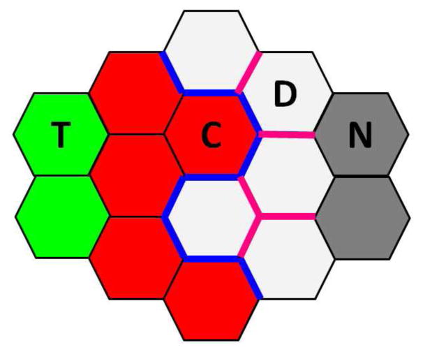
Fig. 2.
Illustration of cell types and mechanical properties within tumor and its microenvironment. Non-invasive tumor cells (T, green) are surrounded by invasive cancer cells (C, red). Invasive cancer cells are at the tumor-host interface, directly degrading the ECM. Degraded ECM (D) are white, and normal ECM (N) far away from the invasive cancer cells are gray. During tumor invasion, tension on the edge between invasive cancer cells and degraded ECM (blue) changes due to the lost adhesion between tumor cells and intension to adhere to the ECM. Tension on the edge among degraded ECM (pink) changes due to degradation by invasive cancer cells and increased stiffness of the ECM.

