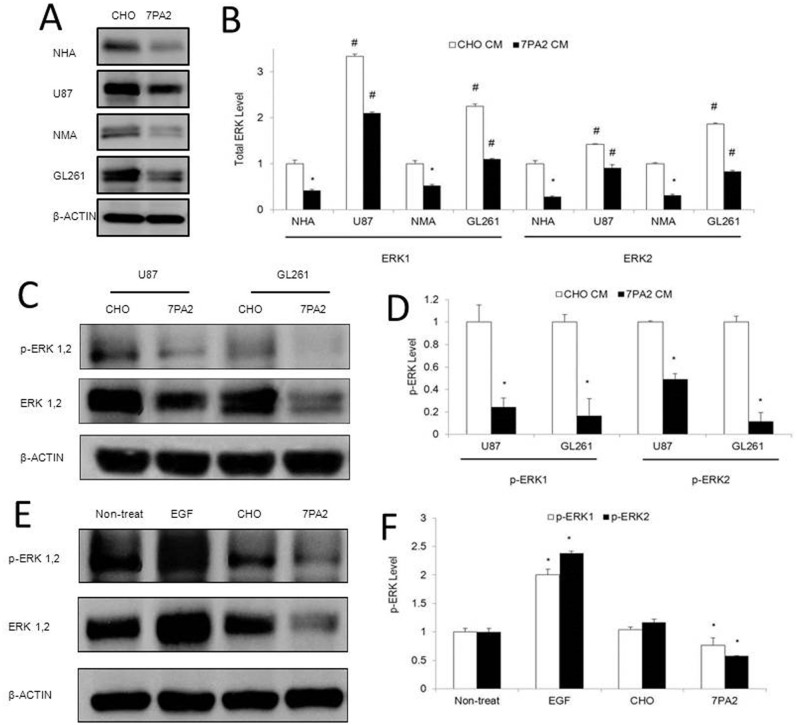
Figure 2. Effects of the Aβ-containing conditioned medium on basal and activated ERK expression in astrocytes and brain tumor cells.
(A–B). Normal human (NHA) and mouse (NMA) primary astrocytes, and human GBM (U87) and mouse GBM (GL261) cells were exposed to Aβ-containing conditioned medium (7PA2-CM) for 72 hours, and total ERK expression was detected by Western blot. (C–D). U87 and GL261 cells exposed to 7PA2-CM for 30 minutes were detected for phosphorylated ERK1/2 (pERK1/2). (E–F). U87VIII cells exposed to EGF and 7PA2-CM for 1 hour were detected for pERK1/2. *P < 0.05 vs. CHO-CM. The quantification data were obtained from three independent experiments under the same condition. In all panels, western blots images shown are cropped to show the protein of interest, and all blots were performed under the same experimental conditions.

