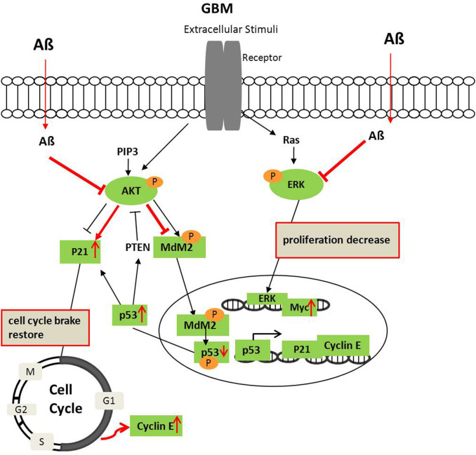
Figure 6. Diagram of Aβ affecting the ERK1/2 and AKT signaling crosstalk in GBM.
AKT and ERK are de-phosphorylated in GBM after Aβ treatment. AKT deactivation decreases MDM2 and p53 phosphorylation and increases stable p53, which trigger its downstream p21 and cyclin E expression. Simultaneously, p53 may increase PTEN to suppress AKT activation further. Red labels indicate the effects of Aβ on GBM.

