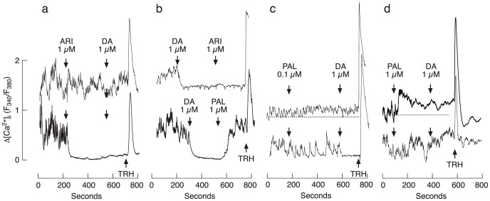
Figure 5. The effect of DA, ARI, and PAL on calcium influx in single rat lactotrophs.
(a) Spontaneous calcium transients were blocked by ARI with no further effect of DA in 19 of 31 cells (bottom trace). In the residual cells, ARI did not abolish spontaneous calcium transients but nevertheless blocked DA-induced inhibition of calcium influx (top trace). (b) Once [Ca2+]i was inhibited by 1 μM DA, 1 μM ARI could not induce any further effect in 14 of 15 lactotrophs (upper trace). 1 μM PAL recovered spontaneous calcium transients previously blocked by DA in 9 of 10 cells (bottom trace). (c) When applied in 0.1 μM concentration, PAL gradually increased [Ca2+]i in a fraction of cells (12 of 48; top trace) and was ineffective in residual cells. In a fraction of non-responders (7 of 48), the subsequent application of 1 μM DA inhibited calcium transients (bottom trace). (d) When applied in 1 μM concentration, PAL rapidly increased [Ca2+]i in a fraction of cells (13 of 45; top trace) and was ineffective in residual cells (bottom trace). In all of these 45 cells, PAL blocked further effect of 1 μM DA. Arrows indicate the moment of drug application, and the subsequent compound was added without dilution of the first compound. Horizontal dotted lines in panels c and d indicate baseline [Ca2+]i.

