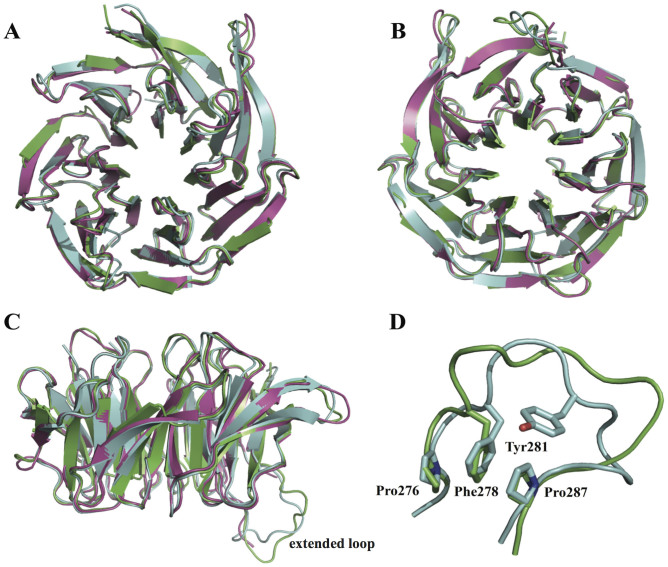
Figure 6. Gib2 comparison with RACK1 and Asc1.
Superimposition of C. neoformans Gib2 in green, human RACK1 (PDB ID 4AOW) in purple, and S. cerevisiae Asc1 (PDB ID 3FRX) in light blue. A, B, and C viewed from the top, bottom, and side, respectively. Close up of the extended loop region (D) is also shown. Side chains of the Asc1 residues involved in the knob-like structure are shown as sticks and labelled individually.

