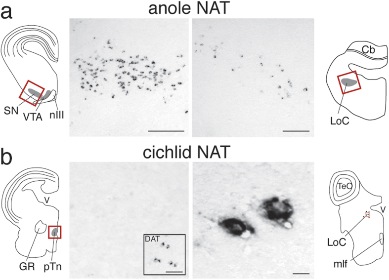
Figure 3. Segregation of NAT and DAT with dopaminergic and noradrenergic cell groups in teleosts, but not reptiles.
(a) In situ hybridization reveals that in green anoles NAT is expressed in the substantia nigra (SN; left) and in the locus coeruleus (LoC; right), consistent with the pattern in finches. (b) In cichlid fish, NAT is expressed in the LoC (b, right), but not in the posterior tuberculum (pTn; left), which contains the teleost equivalent of striatal-projecting dopaminergic cells in VTA/SN. Presence of dopaminergic cells in the pTn is confirmed here by the inset photomicrograph, showing that cells expressing the teleost DAT ortholog are present in the pTn. The locations of photomicrographs are indicated by red rectangles within the camera lucida drawing of transverse brain sections of the green anole (a) and Burtoni cichlid (b) at the level of the midbrain tegmentum (left), and locus coeruleus (right). Anatomical abbreviations: Cb, cerebellum; GR, corpus glomerulosum pars rotunda; LoC, locus coeruleus; mlf, medial longitudinal fascicle; nIII, oculomotor nerve; pTn, posterior tuberculum; SN, substantia nigra; VTA, ventral tegmental area. Scale bars = 100 μm in a; 50 μm in b; 500 μm in b, DAT inset.

