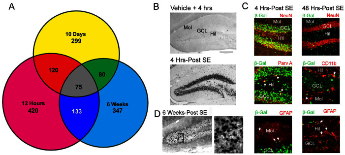
Figure 6. Temporal and cell-type profile of SE-evoked, CREB-mediated, gene expression.
(A) Data from a genome-wide analysis of CREB occupancy by ChIP-Seq was used to determine the proportion of upregulated epileptogenic genes that are CREB-associated. Venn diagram depicts subsets of genes at each timepoint. (B) Representative immunohistochemical labeling of tissue from CRE-β-Gal transgenic mice reveals robust reporter gene expression within the GCL at 4 hrs post-SE onset. Mol: molecular cell layer; GCL: granular cell layer; Hil: hilus. Bar: 50 microns. (C) Double immunofluorescence labeling for β-Gal and NeuN or parvalbumin A (Parv A) revealed that the reporter gene was inducibly expressed in both excitatory and inhibitory neurons at the 4 hour time point. Limited transgene expression was also observed in astrocytes, as assessed via GFAP double labeling. At 2 days post-SE (right panels), limited β-gal was detected in GCL neurons; rather, the reporter was detected in astrocytes and microglia, as assessed via GFAP and CD11b double-labeling. (D) Representative data from animals that were rendered epileptic (sacrificed at 6 weeks post-SE) revealed marked CRE-mediated gene expression within the GCL.

