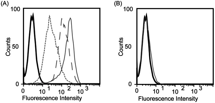
Figure 4. Flow cytometric analysis of T6-17 cells incubated with SPIO nanoparticles.
(A) T6-17 cells were incubated with HER2-SPIO (solid gray line), RGD-SPIO (dashed gray line), and LDS-SPIO (dotted gray line), with varying degrees of cell labeling observed for each ligand. Unlabeled cells are represented by a black solid line. (B) Flow cytometric analysis of T6-17 cells incubated with each variant of the non-targeted LN-doped SPIO nanoparticles.

