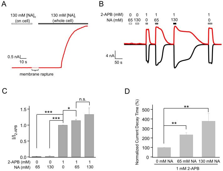
Figure 1. Extracellular nicotinic acid dramatically delays deactivation of 2-APB-induced TRPV1 currents.
(A) Representative current trace from an on-cell recording with 130 mM NA in the pipette followed by membrane rapture, leading to whole-cell patch-clamp recording. (B) Representative whole-cell currents induced by 2-APB and NA applied extracellularly, recorded at +80 mV (top trace) and −80 mV (bottom trace). (C) Comparison of current amplitudes. n = 3–6. (D) Comparison of the slowdown in the deactivation rate, quantified as the time it took for the current to decline to 50% of the peak level. n = 3 to 5. *, p < 0.05; **, p < 0.01; ***, p < 0.005; n.s., not significant.

