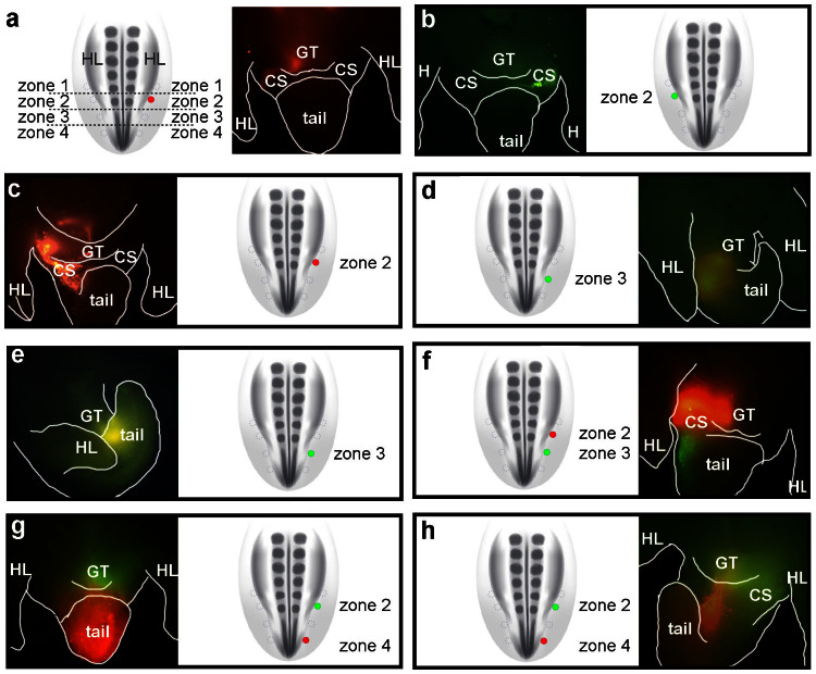
Figure 2. Defining the anteroposterior limits of the external genital progenitor field in the chick lateral mesoderm.
Posterior lateral mesoderm was divided into 4 zones and cells were labeled with DiI or DiA to trace their lineage. In each panel, the schematic diagram is a dorsal view showing the position of dye injection (filled circles) relative to the other zones (empty circles). The adjacent fluorescent micrographs are ventral views showing the distribution of labeled cells 48 hours after injection. (a) Cells labeled with DiI in zone 2 on the right side of the embryo contribute to the right side of the genital tubercle (GT) and the right posterior cloacal swelling (CS). Note that right side of embryo is to the left in ventral views. (b) Cells labeled with DiA in zone 2 on the left side of the embryo contribute to the left side of the GT and the left posterior cloacal swelling. (c) Embryo labeled with DiI on the right side in zone 2. Labeled cells contributed to the right sides of the GT, anterior cloacal swelling, and anterior tail. (d, e) Embryos labeled on the right side with DiA in zone 3. After 48 hours, labeled cells contributed to the posterior part of the GT and the anterior region of the tail (d), or to the anterior region of the tail only (e). (f) Embryo double-labeled on the right side with DiI in zone 2 and DiA in zone 3. After 48 hours, DiI-labeled cells from zone 2 contribute to the right side of the GT and DiA-labeled cells from zone 3 are found posterior to the GT, on the right sides of the posterior cloacal swelling and tail. (g, h) Embryos double-labeled on the right side with DiA in zone 2 and DiI in zone 4. After 48 hours, DiA-labeled cells contribute to the GT and DiI-labeled cells are found in the tail.

