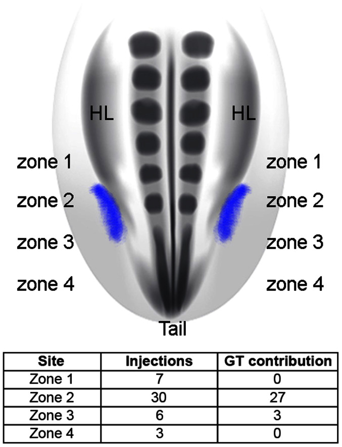
Figure 3. The external genital field maps to the lateralmost mesoderm from the level of the posterior hindlimb bud to the anterior tail bud.
Schematic diagram at top shows the distribution of the external genital progenitor cells (purple) in the chick embryo, dorsal view. The genital tubercle arises from cells situated in zone 2 (posterior hindlimb bud level) and the anterior region of zone 3 (anterior tail bud level). Table shows the results of DiI and DiA injections in each of the 4 zones and their contribution to the genital tubercle (n = 46). Ninety percent of the injections made in zone 2 labeled cells that contribute to the external genitalia. Fifty percent of the injections made in zone 3 labeled cells that contribute to the external genitalia. By contrast, cells labeled in zones 1 and 4 never contributed to the genital tubercle.

