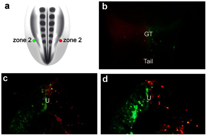
Figure 5. The genital tubercle consists of two lineage-restricted compartments derived from left and right lateral mesoderm.
(a) Schematic diagram of a dorsal view of a stage 18 chick embryo showing the position of two injections in zone 2, one on the right side (DiI, red dot) and one on the left side (DiA, green dot) of the lateralmost lateral plate mesoderm. (b) Ventral view 48 hours after injections. DiI-labeled cells are restricted to the right side (left in ventral view) and DiA-labeled cells are restricted to the left side (right in ventral view) of the genital tubercle. Cells originating from the left and right sides show little to no mixing at the midline of the genital tubercle. (c, d) Transverse sections through the genital tubercle of embryo shown in (b). Descendants of left and right progenitor pools remain restricted to the left and right sides of the genital tubercle. The boundary between the left and right compartments is the urethral plate (U), and neither population crosses into the opposite mesenchymal compartment.

