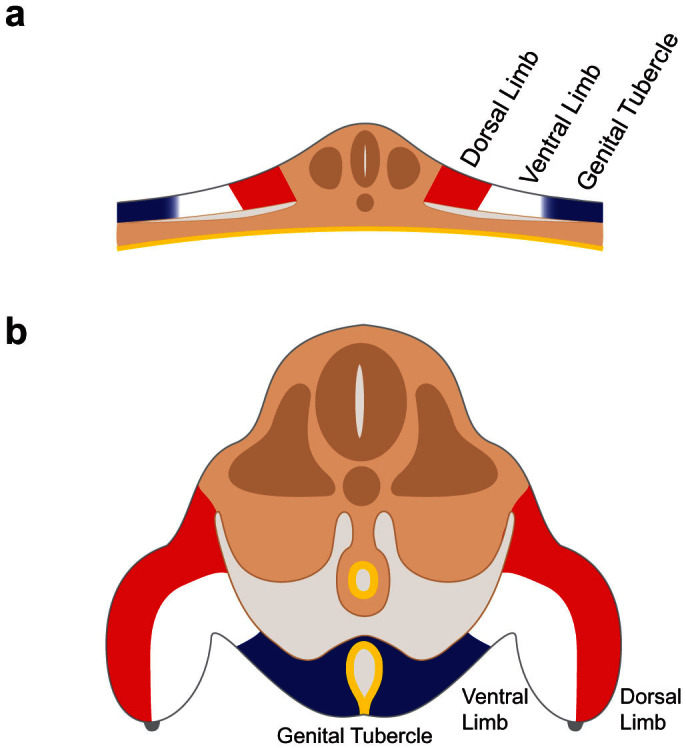
Figure 6. Regionalization of lateral mesoderm into dorsal limb, ventral limb, and external genital fields along the mediolateral axis.

Schematic diagram of transverse sections through the cloacal level of the embryo before (a) and after (b) closure of the body wall. (a) External genital progenitor cells (blue) originate at the lateral edges of the lateral mesoderm, adjacent to the ventral limb field (white). The dorsal limb field is shown in red. (b) Closure of the body wall brings together the left and right external genital fields at the ventral midline, where they give rise to the paired genital swellings that form the genital tubercle (blue). Limb buds are shown on the left and right sides, with the ventral limb shown in white and the dorsal limb shown in red.
