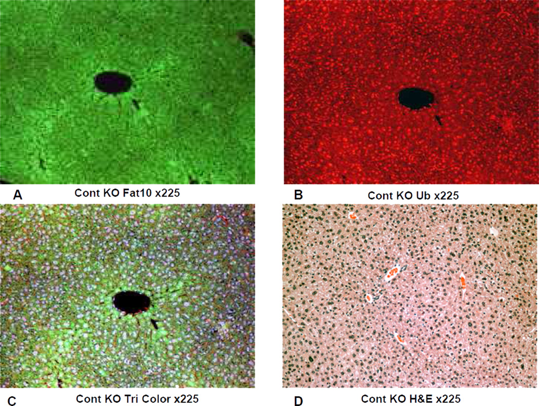
Fig 5.
IHC double stains showing the FAT10 and ubiquitn of a liver from a FAT10 KO mouse fed the control diet without DDC added for 10 weeks. A) FAT10 antibody filter (FITC). B) Ubiquitin antibody filter (Texas red). C) Tricolor filter. D) The same liver stained with Hematoxylin and eosin showing normal liver histology. X225

