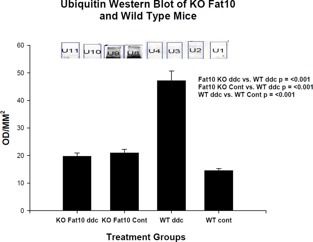
Fig 9.
Ubiquitin smear is present on a Western blot done on the wild type mouse fed DDC for 10 weeks (WT ddc) (U8 and U9). The other 3 groups of mice including the FAT10 KO mice fed DDC (KO FAT10 ddc) did not develop a ubiquitin smear indicating the activity of the 26s proteasome was not diminished in these groups of mice.

