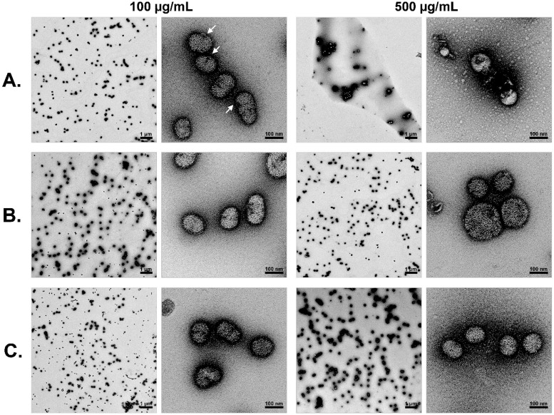
Fig 6. Human BPI-peptide damages the IAV particles.
Virus particles were incubated either with 100 (100) and 500 μg/mL (500) of human (A) or murine BPI-peptides (B) for 1 h or left untreated (C). After the incubation the virus particles were visualized by transmission electron microscopy. Therefore, the particles were negatively stained with 2% uranylacetate and transmission electron microscopy was carried out using a JEOL TEM 2100 at 120kV. Micrographs were recorded with a fast-scan 2k x 2k CCD camera F214. One representative experiment out of 3 performed is displayed.

