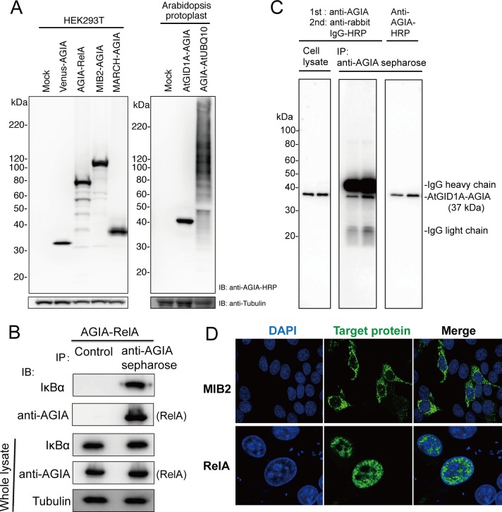
Fig 4. Performance of AGIA tag in Cell biological analysis.
(A) Detection of tagged proteins overexpressed in animal (HEK293T, left panel) or plant cells (Arabidopsis protoplast, right). (B) Co-immunoprecipitation. AGIA-RelA was overexpressed in HEK293T cells (Whole lysate). Immunoprecipitation was performed using anti-AGIA antibody conjugated sepharose (anti-AGIA sepharose). Control experiment was performed using protein G sepharose and normal rabbit IgG (Control). Immunoprecipitated proteins were detected with IκBα specific antibody and anti-AGIA antibody. (C) Performance of anti-AGIA-HRP in immunoprecipitation. AtGID1A-AGIA protein was expressed in Arabidopsis protoplast (left panel). Lysate from protoplast was immunoprecipitated using anti-AGIA antibody sepharose (anti-AGIA sepharose). Subsequently, immunoblotting was conducted using a combination of anti-AGIA and anti-rabbit IgG-HRP antibodies (center) or anti-AGIA-HRP antibody (right), with duplicate sample loading. (D) Immunostaining. MIB2-AGIA and AGIA-RelA were expressed in HeLa cells. Tagged proteins were visualized using anti-AGIA primary antibody with anti-rabbit IgG-Alexa488 secondary antibody (green). Nucleus was stained by DAPI (blue).

