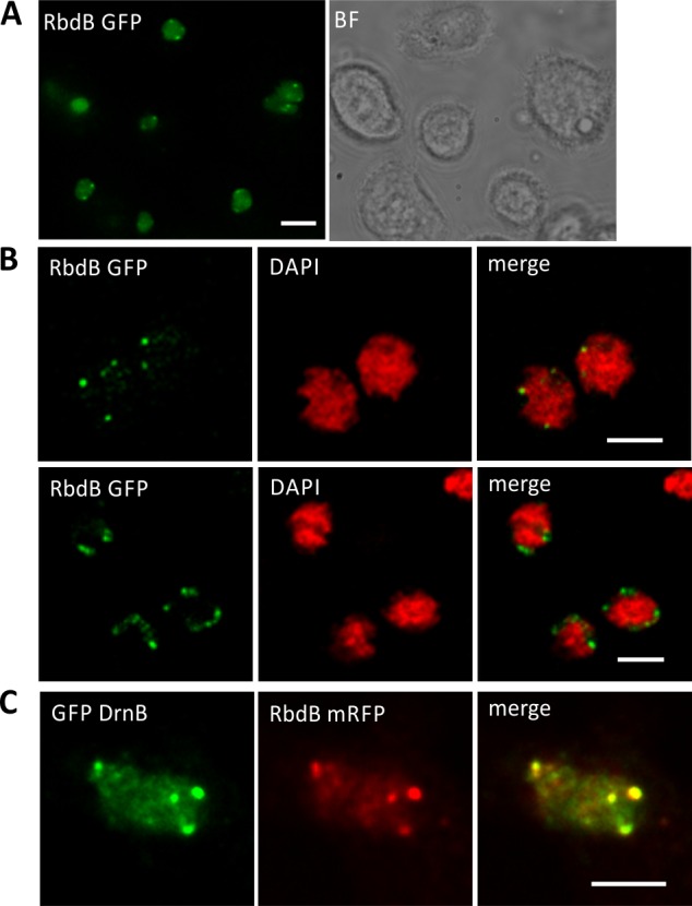
Fig 5. Subcellular localization of RbdB GFP and co-localization with DrnB.

AX2 cells were transformed with the integrating plasmid pDneo2a RbdB GFP and subcellular localization was analyzed by fluorescence microscopy. A: Living cells were analyzed in low fluorescence axenic medium showing a diffuse distribution of the fusion proteins in the nucleoplasm and distinct foci at the periphery of the nuclei. Scale bar represents 5 μm. B: To better localize the subnuclear foci, cells were fixed with methanol and analyzed by an OptiGrid microscope (Leica DM 5500). Genomic DNA was stained by DAPI (red). The nucleoli showed no or only a very weak staining. Merging GFP (green) and DAPI (red) signals indicated that RbdB-GFP foci were enriched adjacent to areas with weak or no DAPI staining. Scale bar represents 2.5 μm. C: Co-localization of GFP DrnB and RbdB mRFP in nucleoli associated foci was monitored by fluorescence microscopy using methanol fixed cells. Shown is a single nucleus. Fusion proteins were expressed from extrachromosomally replicating plasmids. Scale bar represents 2.5 μm.
