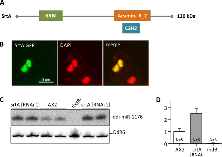
Fig 7. Subcellular localization of the Serrate ortholog (SrtA) in D. discoideum.
A: Protein structure of the D. discoideum Serrate ortholog SrtA. RRM: RNA recognition motif domain, Arsenite-R_2: Arsenite-resistance protein 2, C2H2: Zinc finger domain [43]. B: AX2 cells expressing Srt GFP fusion proteins were fixed with methanol and analyzed by immunofluorescence. DNA was stained by DAPI (red). GFP (green) and DAPI signals were merged. C: ddi-miR-1176 miRNA processing was analyzed in AX2 and in srtA [RNAi 1] and srtA [RNAi 2] knockdown strains. 12 μg total RNA were loaded per lane. Mature ddi-miR-1176 was detected as described in Fig 3A. To show equal loading, the membrane was rehybridized with a probe directed against the snoRNA DdR6. D: The expression level of ddi-miR-1176 was quantified relative to DdR6 and normalized to the AX2 wt. Error bars: mean with SD, paired t-test: ddi-miR-1176: AX2/srtA [RNAi] p < 0, 0001 (***).

