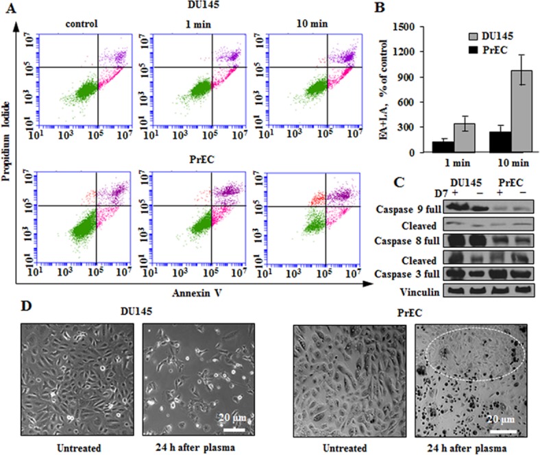
Fig 2. Non-thermal plasma induces apoptosis in DU145 cancer and PrEC normal prostate cells.

Flow cytometry and microscopy results were obtained 24 hours post plasma treatment. (A) Induction of apoptosis in DU145 and PrECs. The cells were incubated with plasma D7 for 1 and 10 minutes than fresh medium was added to cells for their further maintaining. (B) The quantitative data of the per cent of early and late (EA+LA) apoptotic cells. C. Western blot analysis of apoptosis signature. Vinculin was used as a loading control. (D) Transmission images of normal PrEC and metastatic DU145 cells. The white circle indicates the area of PrECs that remained alive or proliferated after the plasma treatment. Data presented as mean±SEM (n = 3).
