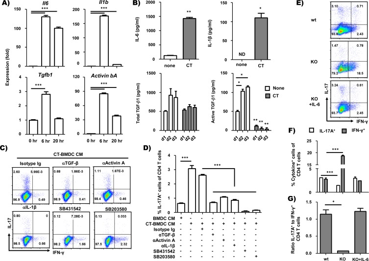
Fig 5. CT–treated BMDCs mediate Th17 cell differentiation by producing Th17 polarizing cytokines.
(A) RT-PCR analysis of IL-6, IL-1β, TGF-β1 and activin A mRNA in BMDCs. BMDCs were cultured with CT (2 μg/ml) and harvested at 0, 6, or 20 h after CT treatment. mRNA levels of IL-6, IL-1β, TGF-β1 and activin βA were normalized by mRNA expression of GAPDH and β-actin. (B) Determination of cytokines in BMDC-conditioned media. BMDCs were cultured with CT (2 μg/ml) for 2 days, and culture media was removed to measure cytokines. IL-6 and IL-1β were assayed by multiplex bead cytokine assay kit following the manufacturer’s recommended protocol (eBioscience). TGF-β1 in BMDC-conditioned media was assayed for total (left) and active form (right) by ELISA (R&D Systems). (C and D) Intracellular staining of IFN-γ and IL-17A in OT-II CD4+ T cells stimulated with anti-CD3 and anti-CD28 antibody in the presence of BMDC-CM or CT-BMDC-CM for 5 days and restimulated with PMA and ionomycin after 5 days. Neutralizing antibodies and kinase inhibitors were also added in the culture as indicated. (E-G) BMDCs from IL-6-/- mice were used for clarifying a role of IL-6 in Th17 differentiation promoted by CT-treated BMDCs. Frequency of IFN-γ+ or IL-17A+ CD4+ T cells (F) and ratio of IL-17+ cells to IFN-γ+ cells (G). *p<0.5, **p<0.01, ***p<0.001 (Student’s t-test). Data are the representative of at least two independent experiments with similar results and average ± SEM of triplicate wells in A and D or duplicate wells in B, F and G.

