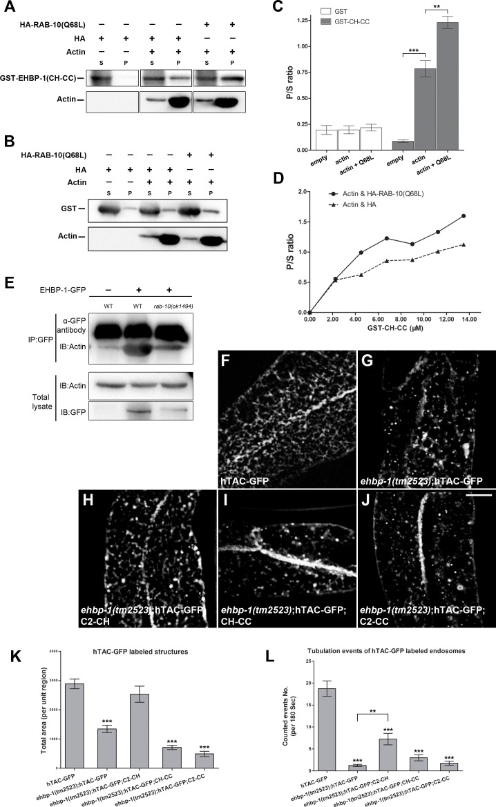
Fig 6. RAB-10(GTP) promotes co-sedimentation of EHBP-1 with F-actin and C2-CH partially restores hTAC-GFP tubular endosomal localization in an ehbp-1(tm2523) mutant background.
(A) GST-CH-CC(260-901aa) co-sediments with actin filaments in vitro. The co-sedimentation level of GST-CH-CC with actin filaments increased by ~67% when complexed with HA-RAB-10(Q68L), a predicted constitutively active form of RAB-10. (B) control protein GST did not co-sediment with actin filaments. P/S ratio (pellet/supernatant) was quantified for GST-CH-CC and GST in (C), error bars are SEM (n = 3), asterisks indicate significant differences in the one-tailed Student’s t-test, ** p<0.01, *** p<0.001). (D) Equilibrium binding of GST-CH-CC to F-actin measured by titrating 21 μM F-actin with 2.25 uM to 13.5 uM GST-CH-CC in the presence of HA-RAB-10(Q68L). (E) Co-immunoprecipitation of EHBP-1-GFP and endogenous actin in wild type and rab-10(ok1494) animals. EHBP-1-GFP was immunoprecipitated with anti-GFP antibody and precipitants were analyzed by immunoblotting using anti-actin antibody. Aliquots of total lysates (2% of the total input into the assay) were examined by immunoblotting using anti-actin and anti-GFP antibodies. (F) In intestinal epithelia hTAC labels basolateral tubular and punctate recycling endosomes. (G) In ehbp-1(tm2523) mutant animals, hTAC-GFP over-accumulated with a significant loss of hTAC-GFP positive tubules. (H) Transgenic expression of the C2-CH fragment partially rescued hTAC-GFP tubular labeling. (I-J) Few tubular structures were observed in ehbp-1(tm2523) mutant intestinal cells with transgenic expression of CH-CC or C2-CC fragments. Total area (per unit region) was quantified for hTAC-GFP labeled structures in (K), error bars are SEM (n = 18 each, 6 animals of each genotype sampled in three different regions of each intestine defined by a 100 x 100 (pixel2) box positioned at random), asterisks indicate significant differences in the one-tailed Student’s t-test *** p<0.001). Scale bar represents 10 μm. (L) Tubule movement events were quantified for hTAC-GFP labeled endosomes in WT or ehbp-1(tm2523) animals as indicated in (F-J). C2-CH expression presented ~7 movement events per 180 sec, compared with ~18 events in wild-type animals and ~1 event in ehbp-1(tm2523) mutant animals. Transgenic expression of CH-CC or C2-CC failed to rescue hTAC-GFP movement defects in ehbp-1(tm2523) mutants. Asterisks indicate significant differences in the one-tailed Student’s t-test (**p< 0.01, *** p< 0.001).

