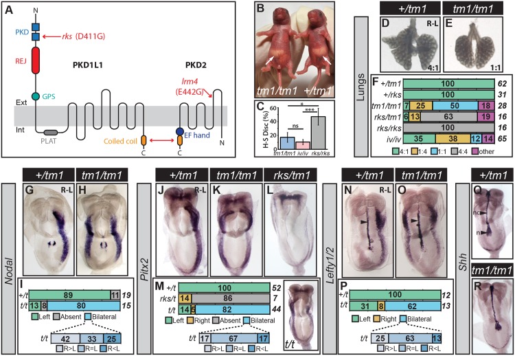
Fig 1. Phenotyping of Pkd1l1tm1/tm1 Mutants.
(A) Schematic diagram of PKD1L1 and PKD2 showing protein domains and the nature of the Pkd1l1rks and Pkd2lrm4 point mutations. The double headed red arrow denotes the site of interaction between PKD1L1 and PKD2. PKD—Polycystic Kidney Disease; REJ—Receptor for Egg Jelly; GPS—G-protein Coupled Receptor Proteolytic Site; PLAT—Polycystin-1, Liopoxygenase, Alpha-Toxin. (B) Pkd1l1tm1/tm1 and sibling control showing reversed and normal situs, respectively. White arrows indicate stomach position. (C) Heart-stomach discordance (H-S Disc.) in Pkd1l1tm1/tm1, Dnah11iv/iv and Pkd1l1rks/rks mutants scored at E13.5. Normally, the heart apex and stomach are positioned to the left. H-S Disc. is defined as the heart apex and stomach being on opposite sides. ns—not significant; *—p<0.05; **—p<0.001, Fisher’s Exact Test applied. (D-F) Lung situs assessed at E13.5 for embryos of the indicated genotypes with the ratio of lung lobes between left and right sides given. The percentage and total numbers of embryos showing each phenotype are indicated in (F). (G-P) Expression patterns of Nodal, Pitx2, and Lefty1/2 in embryos at E8.5 of the indicated genotypes, with the percentage number of embryos exhibiting each phenotype and the total number given. Embryos exhibiting bilateral marker expression are further categorized by whether they show equal or biased expression between the left and right sides. The inset in (M) shows a Pkd1l1tm1/tm1 embryo with bilateral Pitx2 expression but with a right-sided bias. Arrowheads in (N) and (O) indicate midline Lefty1 expression. t is shorthand for Pkd1l1tm1. (Q-R) Sonic hedgehog (Shh) expression in the node (n) and notochord (nc) at E8.5.

