FIG 3 .
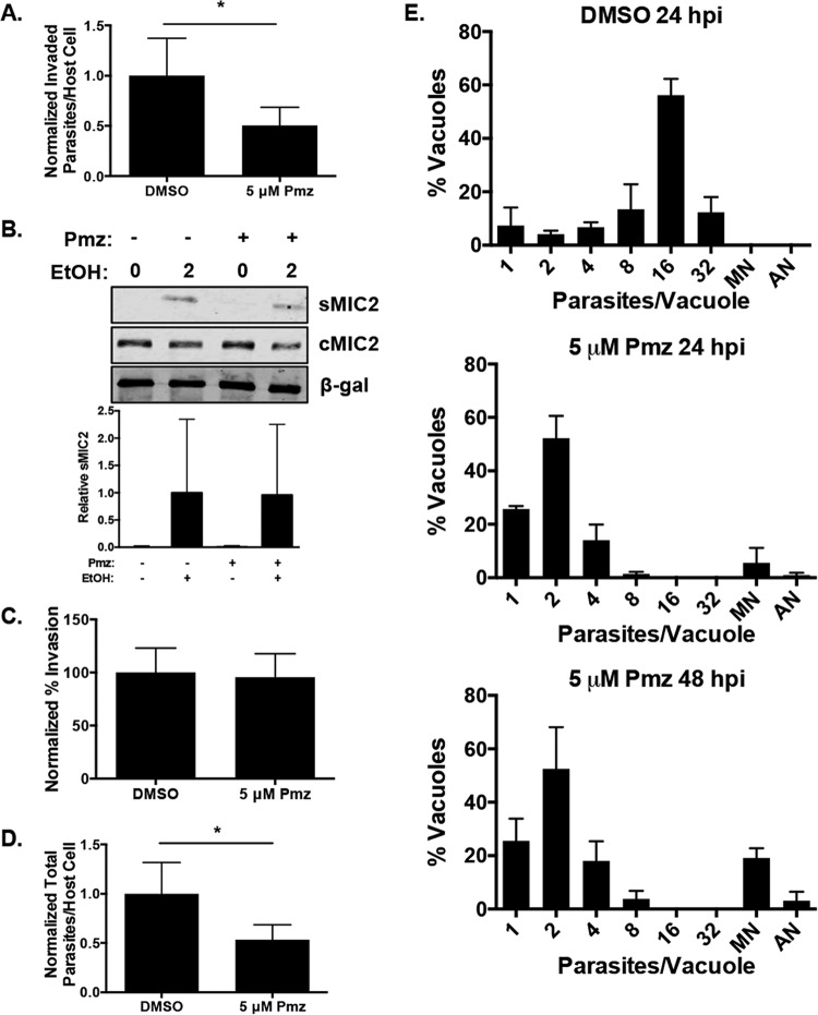
Pimozide inhibits parasite invasion and replication. (A) Host cell monolayers were pretreated with 5 µM pimozide (Pmz) or DMSO for 1 h prior to infection with RH–β-Gal–GFP parasites for 1 h. The cells were fixed without permeabilization and stained with DAPI (to identify host cell nuclei) and anti-SAG1 antiserum. Invasion events were scored via differential staining as GFP+ SAG1− (invaded) and GFP+ SAG1+ (extracellular). A minimum of 500 host cells were counted for each duplicate sample. Shown are the average values and standard deviations from one experiment representative of three independent experiments performed in duplicate. (B) DMSO- or pimozide-treated extracellular RH–β-Gal–GFP tachyzoites were incubated in the absence or presence of 1% ethanol (EtOH) for 2 min at 37°C. The supernatants (sMIC2) and parasites (cMIC2) were Western blotted to detect MIC2. β-Gal was detected as a loading control. Quantification of sMIC from three independent experiments was performed. (C) The percentage of intracellular parasites from panel B was calculated to determine invasion efficiency. (D) The total numbers of parasites per host cell in DMSO- and pimozide-treated cells were compared. (E) RH parasites were allowed to invade HFFs, and 2 h later, DMSO or pimozide was added. After 24 and 48 h, the cells were fixed and stained with anti-SAG1 antiserum and the number of parasites per vacuole was determined. *, P < 0.05 (unpaired Student t test). MN, multinucleated; AN, anucleated.

