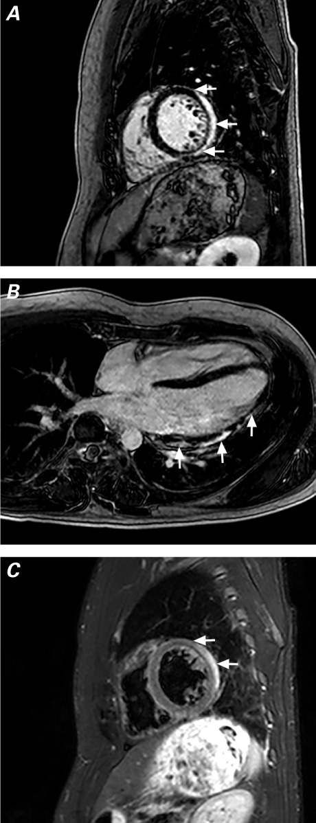
Fig. 1.

T1-weighted cardiac magnetic resonance images acquired during the patient's 2nd hospitalization show A) delayed enhancement (arrows) of the posterolateral wall, sparing the endocardium, and B) heterogeneous delayed enhancement (arrows) of the lateral wall from the midmyocardium to the subepicardium. C) T2-weighted image (corresponding to slice A) shows bright areas of edema (arrows) and a small pericardial effusion.
