
Figure 6.
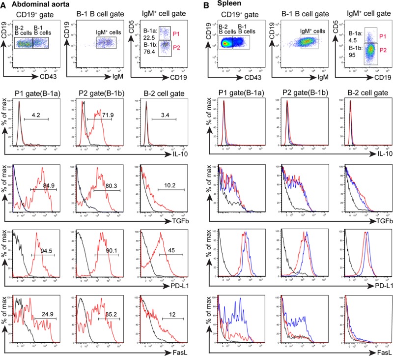
Artery tertiary lymphoid organ (ATLO) B-1 B cells show a predominant immunosuppressive IL-10+/PD-L1+/FasL+/TGFβ+ phenotype. Cell suspensions from individual aged ApoE−/− mice. A, IL-10+, TGFβ+, PD-L1+, and FasL+ abdominal aorta B cells. B, ApoE−/− (red) spleen (80- to 85-week old mice) and WT (blue); ApoE−/− (n=3–4). B-1a, B-1b, and B-2 cell populations were gated and assayed for cytokine expression (or isotype control, black). Numbers designate frequencies of positive cells.