Figure 6.

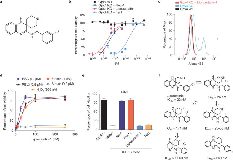
Identification and characterization of ferroptosis inhibitors with in vivo efficacy. (a) Chemical structure of Liproxstatin-1. (b) Dose-dependent rescue of ferroptosis by Liproxstatin-1 in Gxp4−/− cells. On Gpx4 disruption in Pfa1 cells by TAM in the presence of Liproxstatin-1, cell viability was determined 72 h after knockout induction. (c) Liproxstatin-1 (50 nM) completely prevents lipid peroxidation in Gpx4−/− cells. Lipid peroxidation was assessed 48 h after knockout induction using the redox-sensitive dye BODIPY 581/591 C11. (d) Specificity of Liproxstatin-1 towards ferroptosis-inducing triggers. Liproxstatin-1 (200 nM) protects against FINs, such as BSO (10 μM), erastin (1 μM) and RSL3 (0.5 μM), in a dose-dependent manner, whereas it fails to rescue cell death induced by staurosporine (Stauro; 0.2 μM) and H2O2 (200 μM); cell viability was assessed 24 h after treatment. (e) Liproxstatin-1 does not prevent necroptosis. Two hours before triggering necroptosis, L929 cells were treated with Nec1 (10 μM), Nec1s (10 μM), Liproxstatin-1 (1 μM) and Fer1 (1 μM). Necroptosis was induced in L929 cells by a combination of TNFα (5 ng ml−1) and Z-VAD-FMK (50 μM) and cell viability was determined 8 h thereafter using AquaBluer. Data shown represent the mean ± s.d. of n = 4 wells (b,e) or n = 3 wells (d) of a 96-well plate from a representative experiment performed independently at least four times. (f) An initial SAR analysis including IC50 values of Liproxstatin-1 derivatives is shown. IC50 values depicted were calculated from experiments performed with inducible Gpx4−/− cells in 1 well of a 96-well plate, pooled from n=4.
