
Figure 7.
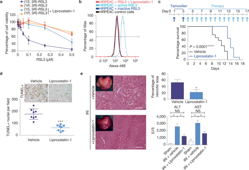
Ferroptosis in human cells and in murine disease models can be targeted by Liproxstatin-1. (a) HRPTEpiCs are susceptible to Gpx4-inhibition-induced ferroptosis, which can be prevented by Liproxstatin-1 (100 nM). Dose-dependent killing of HRPTEpiCs by active (1S,3R)-RSL3 in contrast to inactive (1R,3R)-RSL3. Viability was assessed 24 h after treatment using AquaBluer. Data shown represent the mean ± s.d. of n = 3 wells of a 96-well plate from a representative experiment performed independently three times. (b) Liproxstatin-1 prevents RSL3 (0.2 μM)-induced lipid oxidation in HRPTEpiCs. Lipid peroxidation was assessed 24 h after knockout induction using the redox-sensitive dye BODIPY 581/591 C11. A representative experiment is shown performed independently four times. (c) Liproxstatin-1 retards ARF and death of mice induced by Gpx4 deletion; median survival was calculated to be 11 days for vehicle-treated (n = 12) and 14 days for Liproxstatin-1-treated mice (n = 13), Gehan–Breslow–Wilcoxon test: P <0.0001. Representative experiment shown was performed two times. Mice were injected daily with Liproxstatin-1 (10 mg kg−1, i.p.) during the course of the experiment. (d) Quantification of TUNEL cells in kidneys of vehicle- and Liproxstatin-1-treated animals at 9 +days after TAM administration. Data shown represent the mean ± s.d. of n = 4 comparable anatomical sections from a representative experiment performed two times (scale bars 50 μm). (e) The extent of tissue injury on transient ischaemia/reperfusion in liver of C57BL/6J mice can be ameliorated by the ferroptosis inhibitor Liproxstatin-1 as measured by AST/ALT (n = 17) for vehicle and for Liproxstatin-1 each) and by determining the necrotic area (n = 5). Data represent the mean ± s.e.m.; ∗P = 0.05 or ∗∗∗P = 0.001 (one-way ANOVA) followed by Dunnett’s post-test.