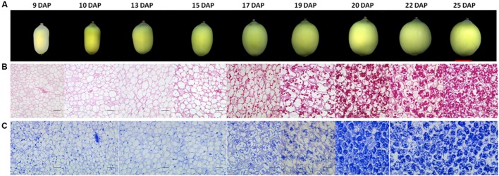
FIGURE 1.
Morphology and histology of developing lotus seeds. (A) Lotus seed at different developmental stages. Histological observation of polysaccharides (B) and protein accumulation (C) during lotus seed development. The polysaccharides and proteins were stained with PAS and Commassie blue method, respectively. The rulers indicate 1 cm in (A) whereas 100 μm in (B,C).

