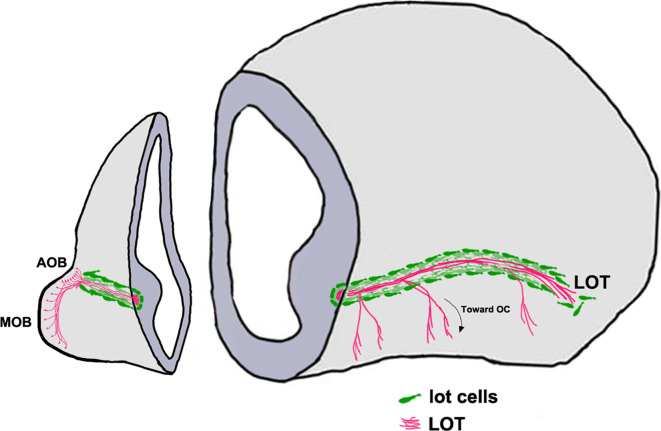
Fig. 4.
“Lot cell” array and formation of the LOT [152, 153]. Schematic representing one hemisphere of an embryonic day (E)14.5 mouse brain. The projection neurons of the MOB and the AOB extend their axons along the LOT and innervate different olfactory cortical and vomeronasal structures. The “lot cells” (green) form a “permissive corridor” along the lateral face of the telencephalon through which the LOT axons (pink) grow. AOB accessory olfactory bulb, LOT lateral olfactory tract, MOB main olfactory bulb

