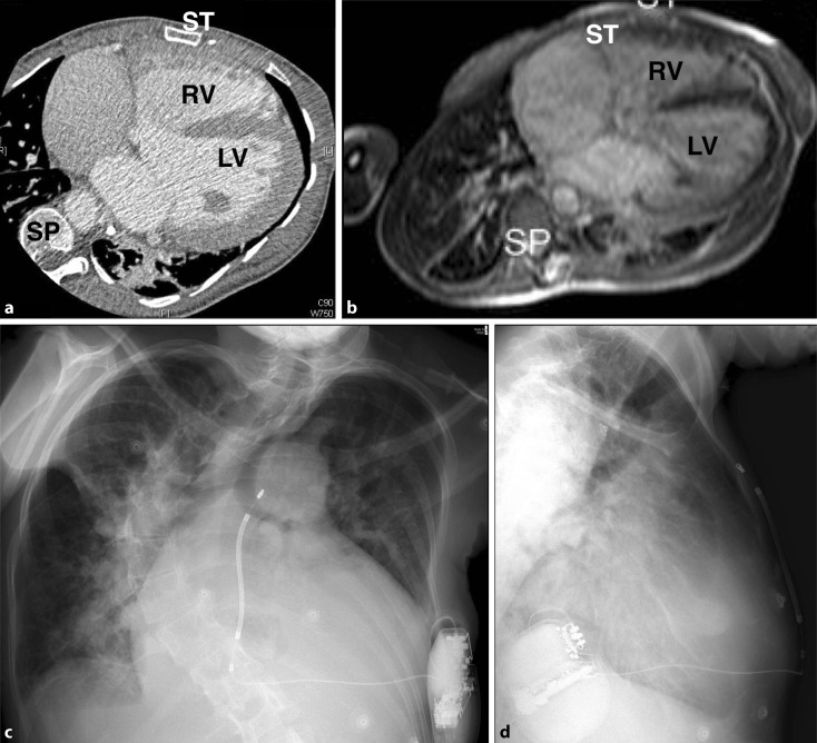
Fig. 4.
Patient with severe scoliosis, pulmonary atresia, ventricular septal defect and major aortopulmonary collateral arteries. a Computed tomography and b Balanced steady state free precession (b‑SSFP) magnetic resonance image. c Anteroposterior and d lateral radiographs demonstrating subcutaneous implantable cardioverter-defibrillator with excellent shock vector (subcutaneous coil has been placed to the right of the sternum, RV right ventricle, LV left ventricle, SP spine, ST sternum)

