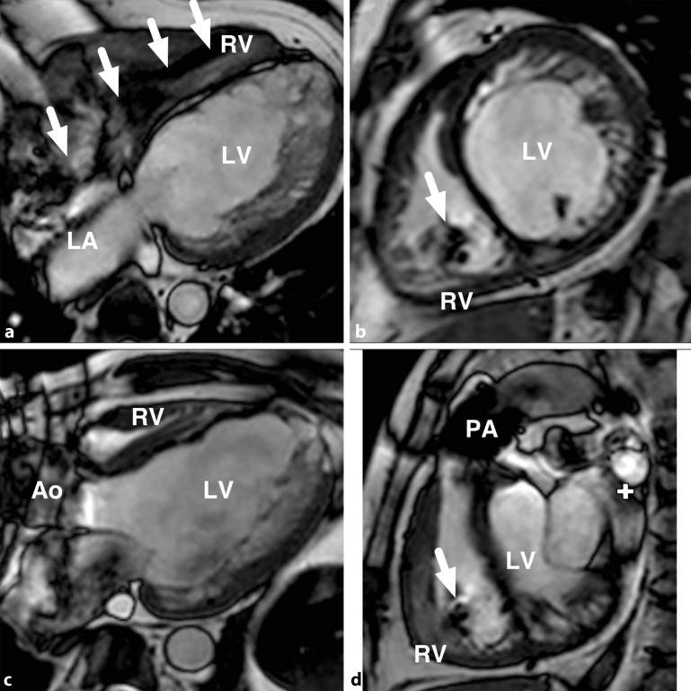
Fig. 6.
Cardiac magnetic resonance imaging in a patient with congenital aortic stenosis (status post-Ross procedure) and magnetic resonance-conditional implantable cardioverter-defibrillator, demonstrating typical results for balance steady state free precession (b‑SSFP) cine imaging. Lead position is indicated by white arrows and the ring artefact related to the generator is seen at the top left of panels (c) and (d). a four chamber view, b short axis, c three chamber view, d right ventricular outflow tract view. RV right ventricle, LV left ventricle, Ao aorta, LA left atrium, PA pulmonary artery

