
Fig. 1.
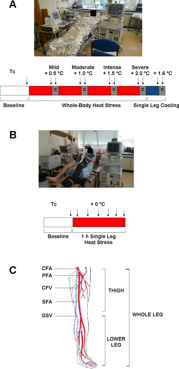
Sequence of the experimental protocols and schematic of the leg major vessels and anatomical sections. A: in study 1, following baseline measurements, participants were heated to core temperature (Tc) + 2°C with measurements (denoted by arrows) taken during mild (+0.5°C), moderate (+1°C), intense (+1.5°C), and severe (+2°C) heat stress, both before and after arterial cuff occlusions (denoted by C) at the level of the knee. Following the whole body heat stress protocol, the left leg was rapidly cooled and further measurements taken as before. B: participants in study 2 rested in ambient thermoneutral conditions while a single leg was heated for a duration of 1 h. C: illustration of the leg's major supplying [common, superficial, and profunda femoral arteries (CFA, SFA, and PFA)] and draining vessels [common femoral vein (CFV) and great saphenous vein (GSV)] and the anatomical differentiation between the thigh and the lower leg.